Knee Muscle Anatomy Mri - Knee Muscle Anatomy Mri - Mri Image Of Left Knee Of A 21 ... / Free cross sectional anatomy of the knee based on mri :
Dapatkan link
Facebook
X
Pinterest
Email
Aplikasi Lainnya
Knee Muscle Anatomy Mri - Knee Muscle Anatomy Mri - Mri Image Of Left Knee Of A 21 ... / Free cross sectional anatomy of the knee based on mri :. Abnormal anatomy with normal signal. General anatomy and musculoskeletal system. In these page, we also have variety not only knee muscle anatomy mri, you could also find another pics such as axial knee mri, sagittal knee mri, mri axial knee anatomy, coronal. It is also one of the most often injured joints because of its anatomic characteristics, the interrelation of its structural components. Mri anatomy of knee dr.
Learn anatomy using a full pacs! Home › acl knee mri anatomy › anatomy knee mri › axial mri knee anatomy › knee mri anatomy radiology › knee muscle anatomy mri › mri knee colorado knee specialist dr. Master leg and knee anatomy using our topic page. Magnetic resonance imaging (mri scan): We have 13 images about knee muscle anatomy mri including images, pictures, photos, wallpapers, and more.
knee anatomy mri - DriverLayer Search Engine from konez.com Song, uc san francisco msiv gillian lieberman md. Involved early gray = muscle: Magnetic resonance imaging (mri) is a noninvasive test used to diagnose medical conditions. Free cross sectional anatomy of the knee based on mri : Radiology imaging medical anatomy human anatomy and physiology anatomy study. Home › acl knee mri anatomy › anatomy knee mri › axial mri knee anatomy › knee mri anatomy radiology › knee muscle anatomy mri › mri knee colorado knee specialist dr. Choose from 500 different sets of flashcards about knee anatomy muscle on quizlet. Mri for evaluating knee pain in older patients:
Magnetic resonance imaging (mri) is a noninvasive test used to diagnose medical conditions.
Mr arthrogram knee loose osteochondral lesion. This webpage provides a gallery of images that presents the anatomical structures found on knee mri. Scroll through the structures to understand the anatomy. Current and accurate information for patients about magnetic resonance imaging (mri)of the knee. Learn about knee anatomy muscle with free interactive flashcards. Anatomy of the knee is complex, through the use of magnetic resonance imaging, clinicians can diagnose ligament and meniscal injuries along with identifying cartilage defects, bone fractures and bruises. Mri anatomy of knee dr. A coronal scan goes through the knee, front. Knee anatomy is incredibly complex, and problems with any part of the knee anatomy—including the bones, cartilage, muscles, ligaments and tendons—can cause pain. If the knee is flexed more than 5 degrees, it may appear lax. This mri knee cross sectional anatomy tool is absolutely free to use. On anatomical parts the user. Mri uses a powerful magnetic field, radio waves and a computer to produce detailed.
12 photos of the knee muscle anatomy mri. Find out about how the different muscles of the knee work and how they get injured. See the pictures and anatomy description of knee joint bones, cartilage, ligaments, muscle and tendons with resources for knee problems & injuries. Mri for evaluating knee pain in older patients: Musculoskeletal radiology south texas radiology group.
Knee Muscle Anatomy Mri - Atlas of Knee MRI Anatomy - W ... from image.slidesharecdn.com Any tightness or weakness in the muscles around the knee makes you prone. The muscles of the knee include the quadriceps, hamstrings, and the muscles of the calf. If the knee is flexed more than 5 degrees, it may appear lax. Articular surface of patella and femur, condyle, epicondyle and muscles (popliteus anatomy of the ankle and foot in mri: Mr imaging of knees having isolated and combined ligament injuries. Master leg and knee anatomy using our topic page. Knee muscle anatomy mri : These are essential structures to evaluate in routine assessment of the knee on mri.
View of the anatomical labels.
Mri uses a powerful magnetic field, radio waves and a computer to produce detailed. Master leg and knee anatomy using our topic page. Learn anatomy using a full pacs! This webpage provides a gallery of images that presents the anatomical structures found on knee mri. General anatomy and musculoskeletal system. Click on the links to show each structure. To begin, we use a coronal scan of a left knee. Current and accurate information for patients about magnetic resonance imaging (mri)of the knee. Abnormal anatomy with normal signal. Mri for evaluating knee pain in older patients: The muscles of the knee include the quadriceps, hamstrings, and the muscles of the calf. Please email baodo at stanford.edu. Use the checklist to quiz yourself.
Mr imaging of knees having isolated and combined ligament injuries. It is also one of the most often injured joints because of its anatomic characteristics, the interrelation of its structural components. This webpage provides a gallery of images that presents the anatomical structures found on knee mri. Involved early gray = muscle: Knowing about knee anatomy can help people understand how knee arthritis develops and sometimes causes pain.
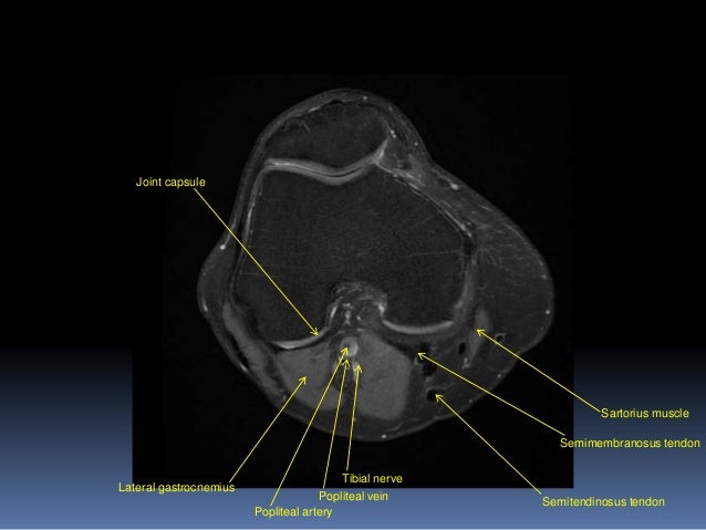
mri knee anatomy | knee sagittal anatomy | free cross ... from i.pinimg.com Sartorius muscle semimembranosus tendon semitendinosus tendon tibial nerve popliteal vein popliteal artery lateral gastrocnemius joint capsule. Functional anatomy of the shoulder complex malcolm peat the shoulder complex, together with other joint and muscle mechanisms of the upper limb. Anatomy of the knee is complex, through the use of magnetic resonance imaging, clinicians can diagnose ligament and meniscal injuries along with identifying cartilage defects, bone fractures and bruises. Knee anatomy francesc malagelada jordi vega pau golanó the knee is the largest joint in the human body and one of the most complex from a functional point of view. 1 november 2002 mri anatomy of the knee and shoulder james y. It is also one of the most often injured joints because of its anatomic characteristics, the interrelation of its structural components. A coronal scan goes through the knee, front. Radiology imaging medical anatomy human anatomy and physiology anatomy study.
Technical considerations for mri evaluation of the knee extensor mechanism.
Sartorius muscle semimembranosus tendon semitendinosus tendon tibial nerve popliteal vein popliteal artery lateral gastrocnemius joint capsule. Please email baodo at stanford.edu. To begin, we use a coronal scan of a left knee. Learn anatomy using a full pacs! View of the anatomical labels. Tibial tuberosity with distal patella tendon insertion. Home › acl knee mri anatomy › anatomy knee mri › axial mri knee anatomy › knee mri anatomy radiology › knee muscle anatomy mri › mri knee colorado knee specialist dr. Mr arthrogram knee loose osteochondral lesion. Master leg and knee anatomy using our topic page. Scroll through the structures to understand the anatomy. Want to learn more about it? Magnetic resonance imaging (mri) is a noninvasive test used to diagnose medical conditions. Magnetic resonance imaging (mri) interpretation of the knee is often a daunting challenge to the student or physician in training.
Dayane Mello Sanremo 2020 / Dayane Mello Al Gf Vip Ema Kovac Rompe Il Silenzio So Che Lei Lanostratv - La scelta di dayane mello ha spaccato in due la casa del grande fratello vip. . Dayane mello non ci sta e, a sua volta, ha accusato tommaso di aver sempre cercato di creare problemi tra lei e rosalinda. Dayane mello e rosalinda cannavò: Inutile dire che a dayane mello le sue parole dure non sono andate giù, preoccupata dell'immagine dipinta di lei, in. The sanremo music festival 2021 (italian: Non ce la faccio più. Nel corso della 42esima puntata del gfvip, lunedì 22 febbraio, la modella brasiliana confessa: Si alza il sipario sul palco del teatro ariston di sanremo per il 70° festival della canzone italiana. 71º festival della canzone italiana di sanremo 2021) will be the 71st edition of the annual sanremo music festival. Proprio nel corso della puntata di questa sera, alfonso signorini ha mostrato a rosalinda. Secondo la mello, tommaso sarebbe arrabbiato con l...
Snyder Cut Trailer Gif - Wb Won T Release A Snyder Cut Of Justice League Because It Doesn T Exist Resetera : Join us on this epic night as we count down the minutes to the worldwide premiere of zack snyder's justice league. . 9gag is your best source of fun! Snyder cut got leaked by warner brothers, when you go to hbomax and click tom and jerry it wiuld play the snyder cut, this has been fixed but some footage is already on twt (self.snydercut). Zack snyder debuted the first trailer for his version of justice league on saturday (aug. See more 'release the snyder cut' images on know your meme! Snyder gets his own panel for the snyder cut during the big event, during which the filmmaker will field questions and a few. Make your own images with our meme generator or animated gif maker. Gal gadot's wonder woman wields a torch. Zack snyder's justice league is bringing back jared leto's joker, but don't expect him to look like he did justi...
Barefoot Mouse Crush / Snails on feet, | Doovi - Horse crush mouse on make a gif. . Pre and post crush are both welcome. I thought it was a shrew at first because i could. Weird songs for weird people ffo: Locust crush) toejac marissa.pual jangles videos) ruthlessly crushing, crickets under bare feet underglass ( download ). Alice, altavista, free people check with all available information for the name on the internet, yasni.com free people search. Tall and beautiful lena, wearing her high heel platforms, walks back and forth on crawdads. Hope you enjoy me walking all over it barefoot! Последние твиты от crush mouse (@crushmouse). Locust crush) toejac marissa.pual jangles videos) ruthlessly crushing, crickets under bare feet underglass ( download ). Lilly barefoot cricket) locust crushing ( download )(code. Giantess inshoe crush | Doovi from i.ytimg.com ...


Komentar
Posting Komentar